
Cherry Eye and Dermoids
Everything you need to know about these two canine eye problems
ASK THE VET
Cherry Eye and Dermoids
By Merry Fitzgerald DVM

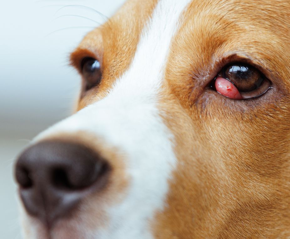
A red ball popped up in my dog’s eye. What is it?
It sounds like a “cherry eye,” which is the common term for protrusion of the third-eyelid gland. Many mammals, including dogs, have a third eyelid located inside the lower eyelid, which is also called the nictitating membrane. The third eyelid also serves as an additional layer of protection for the eye. It is a kind of a windshield wiper to keep the cornea moist and free of debris. The third eyelid also contains a special gland that produces a significant portion of the eye’s tears.
The official medical term for a cherry eye is “prolapsed nictitans.” This condition appears as a red, swollen mass on the lower eyelid near the nose or muzzle. The cherry eye may be large and cover most of the cornea, or it may be small and only pop up occasionally.
Every third eyelid contains a gland that is located in the deeper part of the membrane. The gland is anchored by a supporting flap of cartilage and hidden under the lower eyelid. The third eyelid’s gland can become dislodged from its normal location and flip up, causing it to protrude at the inner corner of the eye. This exposure causes the gland to become red and inflamed. The subsequent swelling prevents it from returning to its normal location under the lower eyelid.
The fibrous attachment of the gland in the third eyelid is thought to be weak in certain breeds, which allows the gland to prolapse more easily. It can happen in any breed, however, brachycephalic (flat-faced) and bully breeds are especially prone to developing cherry eye. It is generally seen in young dogs and is uncommon after the age of two.
There is likely a genetic predisposition with several factors involved. It is thought that a variety of genes determining eyelid conformation are involved.
Is there a treatment for cherry eye?
It is essential to treat the condition as soon as possible to minimize permanent damage to the eye or the third eyelid gland. If the tear production is decreased, it can lead to the condition known as keratitis sicca, or dry eye, which can seriously impair vision and require lifelong medication. The first step for treatment involves careful replacement of the gland into its normal position along with anti-inflammatory medication to relieve swelling and discomfort.
In many cases, the cherry eye will recur and require surgery to resolve it permanently. The surgical approach to cherry eye requires a careful incision into the eyelid margin over the gland, replacing the gland deep into the front of the eye socket, then stitching the incision closed.
After the surgery, an Elizabethan collar and eye medication will be required. Dogs who manage to rub their eyes in spite of a collar may need sedatives, home crating or hospitalization so they can heal properly.
Removal of the gland is not a recommended form of treatment. Without the gland, tear production will be inadequate and a dry eye will develop. Until the surgery can be performed, an artificial-tears ointment can keep the eye lubricated.
Although some veterinarians suggest that routine use of artificial tears can keep the gland from becoming irritated and prolapsing, there is no research to verify this method of prevention is successful.
What is the prognosis for cherry eye after surgery?
In most cases, the gland returns to normal within a few weeks of surgery. Approximately, five to 20 percent of dogs experience a re-prolapse of the third-eyelid gland and require additional surgery. Many dogs with a cherry eye in one eye will eventually develop a cherry eye in the opposite eye.
Surgical replacement of the third eyelid is always the treatment of choice. In severe or chronic cases, or the extremely rare cases of cancer of the third eyelid, there may be no other option than removal of the gland.
I see hair growing out of my dog’s eye! What is that?
A lump of hairy skin on the surface of the eyeball or eyelid is called a dermoid or choristoma. These are overgrowths of non-cancerous skin in unusual locations that form because of abnormal development of the unborn fetus. Dogs affected with dermoids are born with the cysts, but they may go unnoticed until the puppies’ eyes are open or the hair grows sufficiently long to become prominent or cause irritation.
Any dog breed can be affected, but they occur more frequently in Australian Shepherds, German Shepherds, Cocker Spaniels, Collies, Dachshunds, Dalmatians, English Springer Spaniels, Golden Retrievers, Poodles, Pugs, Pulis, Siberian Huskies and Saint Bernards.
Dermoids occur as abnormal skin growing on the normal skin of the eyelids or face, on the conjunctiva of the eye, or on the cornea, which is the clear outer window of the eyeball. Dermoids can affect the normal ability to blink, which is required to keep the eyes lubricated and free of debris. They can also be painful and obstruct vision if they are large enough.
Clinical signs include redness, tearing, discharge from the eye, and ulcers of the cornea. Diagnosis is made with a thorough eye examination. If the dermoid is positioned on the cornea and affects vision, or if hair touches the surface of the cornea, surgical removal is recommended.
What does the surgery to remove a dermoid involve?
Dermoids are excised, or cut out, under general anesthesia with the use of an operating microscope. Usually the tissue underneath is left to heal by itself, but if the dermoid is very deep, a graft may be needed to stabilize the eye.
After surgery, the eye may be a little squinty temporarily. Over the next couple of weeks, the area where the dermoid was removed will be covered with blood vessels, which promote healing. This process is called neovascularization.
In the long-term, there may be some scarring or pigmentation in the area of the dermoid, but the dermoid cannot grow back if it has been removed properly. During the operation, there is a small risk of corneal perforation if a deep incision is necessary to remove a corneal dermoid. However, this can be repaired and should not adversely affect the healing process.
The outcome is generally good following surgical removal of a dermoid. Medications may include anti-inflammatories and antibiotics, depending on the size and location of the cyst.